• Home
  • Facts
  • Peptide Complexes in Ophthalmological Protection: Increased Resilience and Functionality

Peptide Complexes in Ophthalmological Protection: Increased Resilience and Functionality

Introduction

Degenerative changes in young and middle age, as well as age-related changes in the body, are directly associated with the body’s restructuring, psycho-emotional stress, and physical overloads, leading to the development of chronic inflammation. This particularly affects the visual organs. Inflammation is a cellular response to factors that disrupt the homeostasis of cells and tissues. Cell-associated and soluble pattern recognition receptors, such as Toll-like receptors, inflammasome receptors, and components of the complement system, initiate complex cellular cascades by recognizing or sensing various molecular patterns associated with pathogens and damage, respectively. Cytokines and chemokines act as alarm signals for leukocytes, which then travel long distances to the target inflamed tissues. Chronic inflammation is harmful and plays a role in numerous chronic age-related diseases. Degeneration of the retinal pigment epithelium leads to the death of photoreceptors, resulting in the loss of central vision.

The retinal pigment epithelium is susceptible to oxidative stress, a factor that, along with decreased functionality – reduced intracellular recirculation and degradation due to weakened autophagy, leads to a decline in visual function. Clinical medicine is in need of new ways to reduce the progression of degenerative changes, regenerate existing damage, and protect against oxidative stress, inflammation, and destruction. One promising approach to address these issues is the use of peptide complexes. Increasing levels of local and systemic biomarkers released upon peptide application and offering protection indicate the importance of their use in preserving and restoring visual organ functions at any stage of life and in any disease.

Objective: To explore the potential of peptide complexes in ophthalmological protection.

Materials and Methods

This study included 76 participants and was conducted to evaluate the effects of peptide complexes on visual functions, inflammation levels, antioxidant protection, and quality of life over a 3-month period. Participants were divided into two groups: the control group (37 individuals), who received standard ophthalmological care including bi-weekly check-ups by an ophthalmologist, eye exercises, moisturizing treatments (dexpanthenol, hyaluronic acid), and vision correction with glasses; and the main group (39 individuals), who received the same standard care plus the addition of peptide complexes to their treatment regimen.

The peptide treatments were administered according to a specific protocol, starting with Zeaxanthin peptide IPH AVN (4 Vision) for one month, followed by Lutein peptide IPH AVN (4 Vision) for the next month, and concluding with Makui peptide IPH AVN. These German-made peptides (IPH AVN) are fully certified and approved for global distribution, holding credentials such as the WADA (anti-doping) certificate, MAFFA (safety) certificate, ORGANIC certificate, HALAL certificate, and various patents including in the United States (No. 5,405,266), European Union (No. 016704471), Russian Federation (No. 645608), and People’s Republic of China (No. 30507522).

The assessment of the participants involved measuring visual functions, determining levels of inflammation and antioxidant protection, and evaluating the quality of life before the introduction of peptide complexes and after the 3-month period of their application.

Results and Discussion

The study focused on the density of the corneal endothelial layer. In the control group, no significant changes were observed in the layer of posterior epithelial cell count, which was initially 2613.32±7.33 cells/mm² and 2654.36±7.11 cells/mm² after 3 months. However, the main group showed a significant increase in the posterior epithelial cell count, from an initial 2644.32±7.33 cells/mm² to 2771.13±7.2 cells/mm² after 3 months.

Table 1: Dynamics of Cell Density in the Posterior Epithelium of the Corneal Layer (cells/mm², M±m)

Control Group Main Group
Initial Data 3 Months Initial Data 3 Months
Cell Count 2613.32±7.33 2654.36±7.11 2644.32±7.33 2771.13±7.2* **

* p<0.05 compared to initial data, ** p<0.05 compared to data of the control group

This successful increase in the corneal layer is associated with the presence of a patented zeaxanthin formula in the peptide complexes, which counteracts the negative impact of free radicals (r=+0.897, p<0.05), thereby ensuring protection of the lens and retina from damage. Collectively, this enhances high-contrast vision (Figure 1, A), protects the retina from harmful UV radiation (Figure 1, B), shields the retina from photochemical damage (Figure 1, C), and corrects the blind spot area (Figure 1, D).
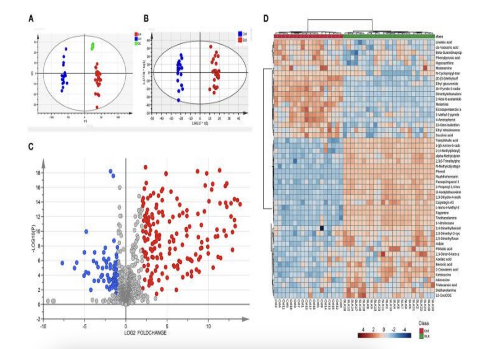
Figure 1: Protective effects of peptide complexes on visual functions.

The incidence of keratopathy signs was examined. It was proven that keratoprotection is only possible with the application of peptide complexes compared to standard preventive measures for the development of eye diseases.

In the main group, after the application of peptide complexes, improvements in keratoprotection occurred by 15%, while in the control group, the improvement was only 10%, and this was only among those patients who initially had mild keratopathies (Table 2).

Table 2: Dynamics of Keratopathy Sign Incidence (Percentage of Patients)

Control Group Main Group
Initial Data 3 Months Initial Data 3 Months
Absent 20% 25% 30% 40%
Mildly Expressed 60% 55% 45% 45%
Moderately Expressed 15% 15% 15% 10%
Significantly Expressed 5% 5% 10% 5%

The keratopathy manifestations, based on their severity, could be categorized into two groups: 1) isolated macular protrusions; 2) macular folds combined with stromal edema, bullous protrusions of the anterior corneal surface in the superficial layers, and decreased corneal transparency.

Following the application of peptide complexes, isolated macular protrusions were observed more frequently, specifically 67.8% more than macular folds with edema. This is associated with the use of the lutein peptide complex, which enhances protection against low-contrast vision (r=+0.881, p<0.05), leading to keratoprotection and improved visual acuity.

Based on the above data, visual acuity was assessed. The data is presented in Table 3.

Table 3: Dynamics of Visual Acuity (Percentage of Patients)

Acuity Detail (min) Control Group Main Group
Initial Data 3 Months Initial Data 3 Months
0.5-0.6 10.0% 5.0% 5.0% 5.0%
0.7-0.8 15.0% 10.0% 15.0% 10.0%
0.9 10.0% 15.0% 10.0% 5.0%
1.0 65.0% 70.0% 70.0% 80.0%

From the table, it is evident that there was a 5.0-15.0% improvement in vision after the application of peptide complexes.

An assessment of the dynamics of dry eye syndrome indicators was also conducted. The data is presented in Table 4.

Table 4: Dynamics of Dry Eye Syndrome Indicators (Percentage of Patients, M±m)

Dry Eye Indicator Control Group Main Group
Initial Data 3 Months Initial Data 3 Months
Schirmer Test<br>(Patients with tear strip wetting < 10mm) 57.0 ± 1.0 66.0 ± 1.2 59.0 ± 1.0 81.0 ± 1.9* **
Norn Test<br>(Patients with tear film break-up time < 10 sec) 61.0 ± 1.0 70.0 ± 1.3* 65.0 ± 1.0 83.0 ± 2.1*

* p<0.05 compared to initial data, ** p<0.05 compared to data of the control group

In evaluating the condition of dry eye syndrome, a significant reduction of 20.0 – 22.0% in dry eye syndrome was observed after the application of peptide complexes.

It is important to note that the degree of restoration of visual function and reduction of dry eye syndrome in patients is associated with the use of the patented peptide complex (Maqui peptide IPH AVN, r=+0.763, p<0.05), which enhances the production of natural tears, mucins, and lipids, affects the evaporation rate of the tear film, and the formation of the tear film on the corneal epithelium surface.

The next stage of the study involved evaluating the reduction in the inflammatory response.

A significant reduction in the tumor necrosis factor-alpha (TNF-α) was noted to be 1.2 times lower after the application of peptide complexes compared to baseline data and data from the control group (Table 5).

Table 5: Dynamics of Inflammation Indicators (pg/ml, M±m)

Study Groups Inflammation Indicators – TNF-α Initial Data 3 Months
Control Group 8.67 + 0.06 8.33 + 0.03
Main Group 8.88 + 0.59 6.52 + 0.52* **

* p<0.05 compared to initial data, ** p<0.05 compared to data of the control group

Upon studying the antioxidant effects following the application of peptide complexes, a 2.2-fold increase in total antioxidant activity was observed, amounting to 496 U/ml in the main group compared to 218 U/ml in the control group. This indicates a strong antioxidant effect of peptide complexes on visual organs.

The data obtained from the use of peptide complexes led to an improvement in quality of life according to the NEI-VFQ (National Eye Institute Visual Function Questionnaire), a survey for ophthalmology.

The analysis of visual functions after the application of peptide complexes improved by 4.5 points, which enhanced the quality of life for patients with medium-level vision impairments to nearly excellent, reaching 24.2 points out of a maximum of 25 points. The primary parameters that patients reported improvements in were: enhanced visual acuity (r=+0.803, p<0.05), color perception (r=+0.791, p<0.05), and reduced dry eye symptoms (r=+0.756, p<0.05).

Thus, the application of peptide complexes leads to a successful increase in the corneal layer, associated with the presence of a patented zeaxanthin formula in the peptide complexes, which counteracts the negative impact of free radicals, thereby ensuring protection of the lens and retina from damage. Collectively, this enhances high-contrast vision, protects the retina from harmful UV radiation, shields the retina from photochemical damage, and corrects the blind spot area. The degree of restoration of visual function and reduction of dry eye syndrome in patients is associated with the use of the patented peptide complex, which improves the production of natural tears, mucins, and lipids, affects the evaporation rate of the tear film, and the formation of the tear film on the corneal epithelium surface. The use of peptide complexes leads to a reduction in inflammation, an increase in antioxidant response, which, collectively, improves the quality of life in terms of enhanced visual acuity, color perception, and reduced dry eye syndrome.


Conclusions

  1. The application of peptide complexes results in a successful increase in the corneal layer, providing protection for the lens and retina from damage, enhancing high-contrast vision, protecting the retina from harmful UV rays, shielding the retina from photochemical damage, and correcting the blind spot area.
  2. The degree of restoration of visual function and reduction of dry eye syndrome in patients is related to the use of the patented peptide complex, which enhances the production of natural tears, mucins, and lipids, influences the evaporation rate of the tear film, and the formation of the tear film on the corneal epithelium surface.
  3. The use of peptide complexes leads to a decrease in inflammation, an increase in antioxidant response, which, collectively, improves the quality of life in terms of improved visual acuity, color perception, and reduced dry eye syndrome.

References

  1.  Ilnickii A.N., Prashchayeu K.I. Neujazvimye. Kniga o zdorov’e [Invulnerable. The book about health]. M.: Diskurs [Discourse]. 2021, 336 p. (In Russian).
  2. Barasheva D. E. Prezhdevremennoe starenie: nenormativnyj krizis identichnosti. Novyj vzgljad [Premature aging: a non-normative identity crisis. New look]. Mezhdunarodnyj nauchnyj vestnik [International scientific Bulletin]. 2016; 12: 109-118. (In Russian).
  3. Ng TP, Nyunt MSZ, Gao Q. Elderly Nutritional Indicators for Geriatric Malnutrition Assessment (ENIGMA): Development and validation of a nutritional prognostic index. Clin Nutr ESPEN. 2017; 22: 54-63.
  4. Thomas CJ, Schroder K. Pattern recognition receptor function in neutrophils. Trends Immunol. 2019; 34: 317–328.
  5. Zong Y, Cheng C, Li K, Xue R, Chen Z, Liu X, Wu K. Metabolomic Alterations in the Tear Fluids of Patients With Superior Limbic Keratoconjunctivitis. Front. Med. 2022; 8: 630-638.
  6. Sinclair D.A., LaPlante M.D. Lifespan: Why We Age―and Why We Don’t Have To Hardcover. New York. 2019, 700 p.
  7. K.I. Prashchayeu, E. E. Sadardinova, M.A. Pokachalova et al. Nutritivnaya podderzhka kak osnova korrekcii prezhdevremennogo stareniya [Nutritional support as a basis for correcting premature aging]. Nauchno-prakticheskij recenziruemyj zhurnal «Sovremennye problemy zdravoohraneniya i medicinskoj statistiki». [Scientific journal «Current problems of health care and medical statistics»].

Related products

Pepts reMuscle

Pepts reVision is the ultimate solution for individuals seeking to improve their eyesight and enhance overall eye health.

Read more

Pepts reMuscle

Pepts reVision is the ultimate solution for individuals seeking to improve their eyesight and enhance overall eye health.

Read more

Pepts reMuscle

Pepts reVision is the ultimate solution for individuals seeking to improve their eyesight and enhance overall eye health.

Read more

Pepts reMuscle

Pepts reVision is the ultimate solution for individuals seeking to improve their eyesight and enhance overall eye health.

Read more

Pepts reMuscle

Pepts reVision is the ultimate solution for individuals seeking to improve their eyesight and enhance overall eye health.

Read more

Pepts reMuscle

Pepts reVision is the ultimate solution for individuals seeking to improve their eyesight and enhance overall eye health.

Read more

Contact us

Explide
Drag