The current global coronavirus pandemic has brought about profound changes at various levels of human life, providing a significant impetus for research into the specific molecular and cellular mechanisms of the body’s response to infection with the new COVID-19 virus, with serotypes that humanity has likely not encountered to such an extent before. It is known that in the human body, several kilograms of biota coexist – microorganisms, viruses, fungi, and in interaction with the microbiota and biotic substances, abiotic substances circulate. Together, this is the internal, relatively harmonized ecological environment of a healthy human being. Coronaviruses, including new serotypes, coexist in the microbiota of a healthy person and have played and continue to play an important role in seasonal outbreaks of acute respiratory viral infections, along with seasonal influenza virus strains. However, according to virologists-infectiologists, other serotypes of coronaviruses have now emerged with which humanity has not yet encountered on its evolutionary path. The appearance of new coronavirus serotypes with new qualities not characteristic of known seasonal pathogen species, in their unpredictable combination with communities of previously formed microorganism ecosystem, as well as against the background of possible chronic infectious and non-infectious pathologies, is accompanied by the development of an individual immune response (humoral or cellular, or both), of different dominant nature and degree of severity [1-3, 20, 21]. Recent studies show a possible dependence on changes in the microbiota of a healthy person after infection with the SARS-CoV-2 virus. Patients with COVID-19 exhibited significant changes in the microbiota compared to the control group, characterized by an enrichment of conditionally pathogenic microorganisms and depletion of beneficial bacteria during illness. The baseline numbers of bacteria Coprobacillus, Clostridium ramosum, and Clostridium hathewayi correlated with the severity of COVID-19, while there was a reverse correlation between the number of Faecalibacterium prausnitzii (anti-inflammatory bacteria) and the severity of the disease [4, 5]. With a high probability, about 80% of cases among those infected with the new COVID-19, can lead to the survival of the affected person and the formation of individual resistance (resistance) after the disease. In turn, at the population level, the pandemic is predicted to end with the formation and preservation of an adaptive result – a collective immune response, which will occur over time. Thus, the problem of the development of a complex COVID-associated pandemic is largely an immunological problem. In this case, immunity acts as the main mechanism and means of protecting the body from genetically foreign substances, viruses, and other infectious agents. The integral immune response that develops in the process of the occurrence and course of coronavirus disease, as it turned out, can proceed unnoticed, at a symptomless level, but can also manifest itself extremely pronounced and life-threatening, in the form of the so-called “cytokine storm.” There is no precise definition and classification description of the “cytokine storm” yet; in a broad sense, it is understood as a hyperinflammatory reaction in which interferons, interleukins, tumor necrosis factors, chemokines, and some other mediators are actively released. The cytokine storm presupposes damage to the body’s own cells due to the release of cytokines and is often fatal [6, 7].
Cytokine release syndrome, or “cytokine storm,” along with acute respiratory distress syndrome (ARDS), lymphopenia, and blood clotting disorders, is one of the key factors in the severe course of the new coronavirus infection COVID-19 caused by the SARSCoV-2 virus [8]. Abnormal elevation of cytokine levels, including interleukin-1β (IL-1β), interleukin-6 (IL-6), interleukin-10 (IL-10), tumor necrosis factor (TNF-α), and vascular endothelial growth factor (VEGF), leads to the formation of a clinical picture of a systemic inflammatory response, in which IL-6 plays a special role [9]. Among all cytokines that increase in COVID-19, IL-6 plays a special role. There is evidence [Cited by 10] of a link between increased levels of IL-6 and a severe course of the disease requiring intensive therapy in COVID-19 patients [11, 12]. Elevated levels of IL-6 in the blood in the context of decreased T-cell immunity increase the risk of death in these patients [13]. A recent meta-analysis of 18 COVID-19 studies, totaling 2984 patients, showed that there is a threshold level of IL-6 in the blood, equal to 1.7 pg/ml, which is a discriminator (from Latin discrimino – “I distinguish”) of mild and severe disease course [14]. With IL-6 levels in the blood below 1.7 pg/ml, the disease proceeded lightly, while exceeding this level led to a severe course of the disease. In the same analysis, it was noted that in non-surviving patients, the level of IL-6 in the blood was above 4.6 pg/ml [14]. There is currently limited understanding of the role of the “cytokine storm” in the severe course of the disease in COVID-19 patients. According to one version [15], high concentrations of cytokines, especially IL-6, negatively affect the survival and proliferation of T cells, which perform the main work of removing virus-infected cells and reducing viral load. High concentrations of IL-6, TNF-α, and IL-10 in serum are significantly correlated with reduced numbers of CD4+ and CD8+ T cells [15, 16], and a decrease in concentrations of IL-6, IL-10, and TNF-α in recovering patients is accompanied by the restoration of the CD4+ and CD8+ T cell pool [15]. Presumably, high concentrations of IL-6 play a role in triggering apoptosis of T cells in the spleen and lymph nodes, which explains the link between the “cytokine storm” and the reduction of the T cell pool in COVID-19 patients [17]. In support of this hypothesis, it is noted that tocilizumab, an IL-6 receptor antagonist, increased the absolute number of circulating lymphocytes in COVID-19 patients during the first 24 hours after administration [18]. Considering the contribution of the “cytokine storm” to the severity and mortality of COVID-19, blocking cytokine signaling systems is considered a potentially effective approach to reducing the severity and lethality of COVID-19.
It should be noted that alongside the successful development of vaccines and effective vaccination, there is still interest in creating new pharmacological agents, with high hopes placed on protein-peptide substances. Specific monoclonal antibodies to interferon-γ (emapalumab), TNF-α (adalimumab), interleukin-6 (siltuximab), and the interleukin-6 receptor (tocilizumab and sarilumab) are currently in clinical trials. However, the cytokine release syndrome involves numerous signaling pathways and a wide spectrum of cytokines. Therefore, despite the high effectiveness of monoclonal antibodies in blocking individual signaling pathways, there is a need for the development of universal agents capable of suppressing or modulating several, and ideally most, signaling pathways that lead to the “cytokine storm” in COVID-19. The activation of the transcriptional nuclear factor kappa B (NF-κB) is a common signaling event that follows the activation of receptors recognizing pathogen-associated molecular patterns, including toll-like receptors TLR3, TLR8, TLR9, and RIGI-like receptors recognizing viral single-stranded RNA (ssRNA) and double-stranded RNA (dsRNA) [11]. NF-κB initiates the transcription of genes encoding cytokines, chemokines, and additional inflammation mediators in various types of innate immune cells. Therefore, NF-κB represents a pharmacological target in the context of suppressing the “cytokine storm” in COVID-19 [10]. Dynorphin 1-17 is a heptadecapeptide from the class of endogenous opioid peptides produced by various cells, including leukocytes. Upon release from leukocytes at the site of inflammation, dynorphin 1-17 undergoes rapid biotransformation, resulting in the formation of a set of fragments. The dynorphin 1-6 fragment has the ability to inhibit NF-κB activation and thus suppress the transcriptional activation of NF-κB-dependent genes, as demonstrated for the cytokines IL-1β and TNF-α [6]. However, dynorphin 1-6 lives less than one minute in vivo due to the action of peptidases, limiting its potential use as a treatment for cytokine release syndrome. As a potential antiviral agent, hexapeptide Leitragin has been proposed, representing a relatively short artificial peptide with an amino acid sequence Tyr-DAla-Gly-Phe-Leu-Arg, corresponding to the structure of the dynorphin 1-6 fragment, in which the Gly residue in the second position is replaced with D-Ala to increase the peptide’s stability to endogenous peptidases [10].
Synthetic hexapeptide, an analogue of leu-enkephalin – tyrosyl-D-alanyl-glycyl-phenylalanyl-leucyl-arginine, known as the drug Dalargin (Tyrosyl-D-alanyl-glycyl-phenylalanyl-leucyl-arginine diacetate), is used in the treatment of gastric and duodenal ulcers during exacerbations and pancreatitis. It inhibits proteolysis and contributes to the healing of ulcers in the stomach and duodenum. It has moderate analgesic action and anti-secretory activity, reducing stomach acid production. In clinical practice, synthetic analogs of endogenous opioid peptides, such as Dalargin (Tyrosyl-D-alanyl-glycyl-phenylalanyl-leucyl-arginine diacetate), are used as anti-stress agents for conditions such as gastric and duodenal ulcers during exacerbations and pancreatitis [22-24]. This hexapeptide, as a non-selective agonist of µ- and δ-opioid receptors, has a wide range of biological activities (analgesic, immunomodulatory, antioxidant, etc.) [25-32]. Similar to an antiviral agent, opioid oligopeptides such as Orvidal and others are being investigated.
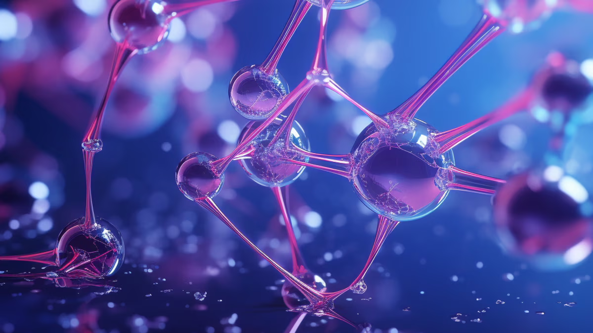
It has been established that the molecular target of SARS-CoV-2 or the entry point for the COVID virus is the angiotensin-converting enzyme 2 (ACE2) receptors located on the surface of lung alveolar epithelial cells. ACE2 regulates fluid and electrolyte exchange in lung tissue [33, 34].
Currently, drugs are being developed that can block this receptor and prevent the virus from interacting with it, thus preventing it from entering cells. In China, there are already trials of such drugs based on oligo- and polypeptides – short-chain proteins. In addition, a class of peptide compounds and possibly nucleoproteins of natural origin, known as Transfer Factors (TF) or Transfer Factors (TF), is considered as promising candidates [35]. The structure of transfer factors is actively being studied, but it remains unknown – some studies indicate a polypeptide nature of TF, while others show sensitivity to certain proteolytic enzymes and RNAase, which cleaves double-stranded RNA, suggesting a nucleoprotein nature of TF. There is a well-founded opinion that transfer factors can be classified as cytokines. Studied TFs are peptides consisting of 44 amino acids with an average mass of 5 kDa. It is claimed that these molecules, numbering more than a hundred, are biochemically and physiologically universal for all vertebrate animals, but the combination of amino acids in their composition determines the specificity of the information encoded in them. Transfer Factors are still insufficiently studied, and recently they have been classified as cytokines, with the role of information transfer agents within the immune system, like all cytokines. Transfer Factors are likely to be peptide multifunctional molecules synthesized within the immune system itself for its integral tuning, control, and intercellular communication. It is assumed that the original amino acid sequence embedded in the structure of oligo- or polypeptides of TF encodes specific signaling information important for the formation of a particular immune response. In the molecular diversity of transfer factors, three fractions of signaling molecules are distinguished by their functional properties: inductors, suppressors, and antigen-specific transfer factors, performing amplifying, inhibitory, and informational functions in the process of forming an immune response. Thus, according to modern views, transfer factors are the main modulators of the links of the body’s immune system, the mechanisms of action of which are actively studied.
As of today, Transfer Factors are considered dietary supplements (nutritional supplements) and are not classified as pharmaceuticals. However, dietary supplements based on transfer factors are approved for adjunctive treatment of diseases such as hepatitis B and diabetes. Several dietary supplements of both imported and domestic manufacture containing TF are widely used in medical practice and in the menu of sports and functional nutrition. In this regard, it is important to note that the term “Transfer Factor” is not appropriate to propose as the name of a specific pharmaceutical product (drug), dietary supplement, or trademark, as the phrase “Transfer Factors” refers to a scientific term denoting a relatively large class of substances of peptide nature, rather than any individual substance, ingredient (e.g., food), or brand name. Currently, scientific classification tends to define TF as substances of peptide (oligopeptides, polypeptides, proteins) nature with a certain range of molecular mass, number of amino acid residues, and spatial configuration. However, further comprehensive experimental physical, biochemical, and physico-chemical studies of transfer factors do not exclude the possibility of expanding the understanding of their molecular composition, such as nucleoprotein nature, in the case of human transfer factors.
Pepts reVision is the ultimate solution for individuals seeking to improve their eyesight and enhance overall eye health.
Pepts reVision is the ultimate solution for individuals seeking to improve their eyesight and enhance overall eye health.
Pepts reVision is the ultimate solution for individuals seeking to improve their eyesight and enhance overall eye health.
Pepts reVision is the ultimate solution for individuals seeking to improve their eyesight and enhance overall eye health.
Pepts reVision is the ultimate solution for individuals seeking to improve their eyesight and enhance overall eye health.
Pepts reVision is the ultimate solution for individuals seeking to improve their eyesight and enhance overall eye health.