• Home
  • Research
  • IPH Peptide Complexes for Treating Ophthalmic Conditions

IPH Peptide Complexes for Treating Ophthalmic Conditions

Introduction

In today’s pharmaceutical market, biological drugs have emerged as one of the most promising avenues. The biopharmaceutical sector offers significant advantages, such as rapid and efficient production capacity utilization and the development of safer and more effective medications. Biological drugs (BDs) differ fundamentally from synthetic substances, as they involve the use of living cells in their production. Each production cycle results in a unique pharmaceutical product, and even minor variations in production methods can significantly impact a drug’s properties.

Preserving the properties and quality of biological drugs at every stage of handling is a current challenge in the pharmaceutical market. Efforts are actively underway to address these challenges, including the development and implementation of quality control systems according to international standards. The aim is to maintain the maximum effectiveness of biological drugs and protect consumers from subpar products.

In ophthalmology, IPH peptides for treating ophthalmic conditions have been in use since 2015. Keywords: ophthalmology, peptide complexes, vision diagnostics, IPH short peptides.

Targeted Treatment and General Information

It’s worth noting that there have been relatively few studies on targeted treatment of ophthalmic diseases using peptides and their complexes. This represents a relatively new alternative approach to anti-angiogenic therapy. It’s important to highlight that targeted anti-angiogenic therapy strategies are relevant for treating ophthalmic diseases associated with pathological neovascularization, as well as for blocking neovascularization in malignant tumors [3].

There are generally two approaches to blocking angiogenesis processes in targeted anti-angiogenic therapy. In one case, immunotoxins are directed at ligands that stimulate angiogenesis (angiogenic factors), while in the other, they target endothelial cell receptors responsible for activating the kinase cascade that drives cell proliferation. When immunotoxins act on ligands, the conjugated antibodies accumulate in the extracellular matrix, and the photodynamic action primarily targets endothelial plasma. In this scenario, the effectiveness of anti-angiogenic therapy is somewhat reduced [3]. When the phototoxin targets endothelial cell receptors, there is an opportunity for internalization of the photosensitizer into the cell. In this case, endothelial cells are more likely to undergo apoptosis, thus increasing therapy’s effectiveness. It’s important to note that antibodies and liposomes are most commonly used as carriers for targeted anti-angiogenic photodynamic therapy [2].

Currently, a range of monoclonal antibodies specific to VEGF or its receptors has been developed. Some of them, when conjugated with photosensitizers, have shown promising results in ophthalmology [4]. There is also information about the use of liposomes conjugated with photosensitizers in targeted anti-angiogenic photodynamic therapy [5].

Clinical Trials on Mice

Experiments on mice with transplanted Colon 26 NL-17 carcinoma and Meth-A sarcoma showed that liposomes conjugated according to the scheme liposome-benzoporphyrin-APRPG-polyethylene glycol have significantly better selectivity in accumulation compared to liposomal conjugate APRPG-polyethylene glycol-liposome-benzoporphyrin and polyethylene glycol-liposome-benzoporphyrin, which, in fact, cannot orient themselves to cells [6]. This is why tumor growth inhibition was noted in this experiment, indicating that photodynamic therapy was directed specifically at damaging endothelial cells and proved to be more effective. Radiation was conducted after 3 hours, as it was believed that within this time frame, the conjugates would better bind to endothelial cells, although some authors suggest that the conjugate is distributed within endothelium as early as 15 minutes after administration [7]. Furthermore, it was found that 3 hours after photodynamic therapy, vascular occlusion and disturbances in the hemostatic system occurred [6].

Significant results have been obtained when blocking pathological angiogenesis in the retina using a conjugate of verteporfin-derivative Bpf with polyvinyl alcohol and the peptide ATWLPPR. This conjugate exhibited high affinity for the VEGFR2 receptor of vascular endothelial growth factor (VEGF). The ratio of verteporfin to peptide in the conjugate was 28:1, while all physicochemical properties of the sensitizer were preserved. On histological sections of the retina after photodynamic therapy, necrotic processes in the endothelium were observed within an hour [7]. The results obtained in ophthalmology indicate an effect on the vascular system, allowing for the application of this approach in similar conditions.

Clinical Trials on Rabbits

In experiments involving a rabbit model of eye angiogenesis, a conjugate with indocyanine of the McATscFvL19 fragment, specific to fibronectin—one of the main markers of the vascular network—also showed a blockade of neovascularization processes. However, this conjugate exhibited significant drawbacks due to its low singlet oxygen production with the photosensitizer indocyanine [9].

When it comes to research on immunotoxins targeting the VEGF ligand, it’s worth noting that there is no such research available. Recently, a drug based on conjugated verteporfin with Visudyne—an antibody to the VEGF factor—has emerged [10]. When determining cytotoxicity, it was found that conjugated verteporfin is more toxic than the free form, although there was no statistically significant difference between them. Thus, conjugated verteporfin with Visudyne has the potential to be an effective drug in photodynamic targeted anti-angiogenic therapy.

It can be assumed that the effectiveness of antibody-conjugated drugs aimed at blocking VEGF activity may be explained not only by photodynamic action but also by neutralizing the action of the VEGF pool in the extracellular matrix. This prevents the factor from binding to VEGFR receptors, thereby inhibiting the proliferative signal of endothelial cells [10]. This to some extent can compensate for the inability of the photosensitizer to internalize into the cell, where photodynamic action would be significantly more effective. Moreover, by targeting the photoimmunoconjugate at VEGFR receptors, a similar synergistic effect can be achieved, as binding of the photoimmunoconjugate to the receptor can block its function, resulting in the inhibition of endothelial cell division.

Direct Treatment of Eye Diseases in Rabbits

In another experimental study conducted in 2015, 66 sexually mature Chinchilla breed male rabbits weighing 3.5 – 5 kg were used. These rabbits were kept on a vivarium diet during the experiment.

The rabbits were divided into the following groups:

  1. Group 1 – Intact (6 rabbits).
  2. Group 2 – Experimental – rabbits with experimental retinal vascular thrombosis (12 rabbits).
  3. Group 3 – Traditional drug therapy – rabbits with experimental retinal vascular thrombosis treated with traditional medications (6 rabbits).
  4. Group 4 – Research – rabbits with experimental retinal vascular thrombosis, where the optimal therapeutic concentration of a polypeptide drug was studied to achieve a therapeutic effect (15 rabbits).
  5. Group 5 – Traditional therapy – rabbits with experimental retinal vascular thrombosis treated with a polypeptide drug at a dose of 0.12 mg/kg intramuscularly for 10 days (6 rabbits).
  6. Group 6 – Traditional therapy – rabbits with experimental retinal vascular thrombosis treated with a polypeptide drug at a dose of 0.5 ml subcutaneously for 10 days (6 rabbits).
  7. Group 7 – Laser – rabbits with experimental retinal vascular thrombosis treated with laser therapy using a selective laser technique on the pigmented retinal epithelium alongside traditional drug therapy (15 rabbits) [12].

A technique was developed for reproducing experimental retinal vein thrombosis, involving a combination of affecting the hemocoagulation potential with thrombin and directly targeting the vascular endothelium using retinal laser coagulation. This ensured the absence of vessel and retinal tissue trauma and provided a sufficiently high and reproducible thrombosis [12].

Central retinal vein thrombosis (CRVO) was reproduced by applying argon laser coagulation to both retinal arteries and veins on both sides of the optic nerve disc at intervals of 1.0 hour. 40 units of standard thrombin from a sodium chloride solution were introduced 30 minutes before coagulation. Argon laser coagulation of retinal vessels was performed using a solid-state laser with a power of 0.4 – 0.8 mW, a coagulation diameter of 50 – 100 μm, an exposure time of 0.2 – 0.3 seconds, and a wavelength of 532 nm [13].

For local anesthesia, a 2% solution of dicaine was used. Premedication included 1 ml of 1% dimedrol solution and 3 ml of 5% analgin solution administered intramuscularly. Hexenal (2 ml of a 5% solution) was injected intraperitoneally for anesthesia. Atropine sulfate (0.1% solution) or tropicamide (1% solution) was used for medicated mydriasis. Direct ophthalmoscopy, examination, and photorecording of the fundus image were performed using a “Carl Zeiss” retinal camera. Fluorescein angiography was carried out using the FF 450 fundus camera by “Carl Zeiss.” Euthanasia was performed under intravenous thiopental sodium anesthesia after the disappearance of the corneal reflex. Laparotomy with caudal vena cava incision and bloodletting was performed afterward. Blood, tear fluid, and eye tissue were also analyzed [13].

For histological examination, the animal eye specimens were cut into two halves and placed in a 10% neutral formalin solution for 5 days.

The first series of experimental research focused on creating an experimental model of retinal vein thrombosis and studying ophthalmoscopic, fluorescein angiographic, histological, and electron microscopic changes in rabbit retinal blood vessels under thrombosis conditions.

The second series of experimental research aimed to determine the optimal concentration for use in retinal vein thrombosis conditions. Concentrations of 0.01, 0.05, 0.12, 0.5, and 1.0 µg/ml were studied. The drug was administered once a day subcutaneously into the right eye of the rabbit. Isotonic sodium chloride solution was administered to the left eye of the animals. Clinical observation included biomicroscopy and direct ophthalmoscopy [13].

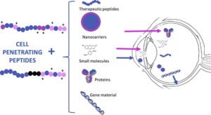
Figure 1. Influence of Peptides on the Retina and Eye

Peptide drugs belong to the group of short peptides found in the structural formations of peptide-binding proteins of the major histocompatibility complex and molecular chaperones. A complex of natural peptides with a molecular mass of 10 kDa has a pronounced anti-exudative and collagen-protective effect.

The third series of experimental research focused on studying the influence of the polypeptide drug on eye structures under physiological conditions. The drug was administered to the intact group of rabbits subcutaneously daily for 10 days at a dose of 0.12 µg/ml.

The fourth series of experimental research aimed to study the therapeutic effects of doses of 0.12 mg/kg and 0.5 ml subcutaneously on the course of the thrombotic process, microcirculatory, coagulation hemostasis, lipid peroxidation, and morphological changes in the eye tissues of rabbits with experimental retinal vascular thrombosis.

The fifth series of experimental research was dedicated to studying the clinical course of the thrombotic process and morphological changes in eye tissues under conditions of experimental thrombosis and treatment with traditional medications and selective laser coagulation of the pigmented retinal epithelium.

Traditional drug therapy included the administration of direct-acting anticoagulant heparin at 250 units and fibrinolysin at 0.3 ml subcutaneously daily for 10 days.

Rabbits with experimental retinal vein thrombosis who underwent selective laser coagulation of the pigmented retinal epithelium were divided into two subgroups. The first subgroup consisted of 9 rabbits who received selective laser coagulation of the pigmented retinal epithelium 7 days after the clinical signs of retinal vein thrombosis appeared. The second subgroup consisted of 6 rabbits who underwent the same laser treatment 14 days after the clinical signs of the disease appeared. The first subgroup of rabbits received focal coagulation of the central retinal area (3 rabbits), coagulation of ischemic areas of the retina (3 rabbits), and panretinal coagulation (3 rabbits). The second subgroup of rabbits underwent focal coagulation of the central retinal area (3 rabbits) and panretinal coagulation (3 rabbits). Euthanasia with subsequent enucleation of the eyes and histological and electron microscopic studies were conducted at different times: for the animals in the first subgroup – on the 10th (3 rabbits) and 30th (3 rabbits) days from the onset of clinical retinal vein thrombosis; for the animals in the second subgroup – on the 20th (2 rabbits) and 30th (2 rabbits) days from the onset of clinical signs of the disease. Observations for 3 rabbits from the first subgroup and 2 rabbits from the second subgroup continued for up to three months. The effectiveness of the peptide complex for treating these diseases was confirmed [13].

Clinical Studies on Retinal Venous Circulation Disorders

Clinical studies were conducted on 232 patients with acute disorders of retinal venous circulation. The age of the patients ranged from 35 to 78 years. The participants were divided into the following groups:

  1. Control Group: 30 individuals without acute retinal venous circulation disorders.
  2. Group 2: 52 patients with acute retinal venous circulation disorders who received traditional medication therapy, including heparin and aspirin.
  3. Group 3: 30 patients with acute retinal venous circulation disorders who received traditional medication and, when necessary, laser treatment.
  4. Group 4: 30 patients with acute retinal venous circulation disorders who were treated with traditional methods and the anticoagulant, enoxaparin.
  5. Group 5: 50 patients with acute retinal venous circulation disorders who were treated with traditional methods. This group used antiplatelet agents, either ticlopidine or clopidogrel.
  6. Group 6: 30 patients with acute retinal venous circulation disorders who were treated with traditional methods, as well as a polypeptide bioregulator.
  7. Group 7: 20 patients with acute retinal venous circulation disorders who received selective laser coagulation of the retinal pigment epithelium in addition to traditional medication therapy.
  8. Group 8: 20 patients with acute retinal venous circulation disorders who received comprehensive treatment, including enoxaparin as an anticoagulant, ticlopidine or clopidogrel as antiplatelet agents, a polypeptide preparation, selective laser coagulation of the retinal pigment epithelium, as well as fibrinolytics, antihypertensive drugs, and corticosteroids.

In Group 2, patients were treated with heparin and aspirin. In Group 4, enoxaparin was administered daily, and in Group 5, antiplatelet agents such as ticlopidine or clopidogrel were used. Group 6 received a polypeptide bioregulator, and Group 7 underwent selective laser coagulation of the retinal pigment epithelium in addition to traditional medication therapy. Group 8 received a comprehensive treatment regimen that included enoxaparin as an anticoagulant, antiplatelet agents (ticlopidine or clopidogrel), a polypeptide preparation, selective laser coagulation of the retinal pigment epithelium, fibrinolytics, antihypertensive drugs, and corticosteroids. The results of using peptides in this study showed high effectiveness.

Chororetinal Dystrophies

In recent decades, there has been a sharp increase in the proportion of chororetinal dystrophies among eye diseases, with age-related macular degeneration (AMD) taking a leading position among them. AMD is also known by other names such as senile macular degeneration, sclerotic disciform degeneration, age-related macular dystrophy, central involutional retinal dystrophy, Kunt-Junius dystrophy, and others.

Currently, there is research that establishes a connection between the consumption of long-chain omega-3 fatty acids and the progression of AMD, particularly the development of the wet form. Clinical study results, such as those from the AREDS study, suggest that consuming high doses of omega-3 fatty acids, especially docosahexaenoic acid (DHA), along with food, reduces the risk of developing the wet form of AMD by 46%. Omega-3 fatty acids are known for their anti-inflammatory properties, prevention of pathological neovascularization, and improvement of blood rheological properties. DHA, in particular, plays a crucial structural role, comprising 56% of the outer membranes of photoreceptors.

Modern approaches to the pharmacological treatment of subretinal neovascularization are somewhat contradictory. Conservative treatment of transudative macular degenerations, involving local administration of glucocorticoids and various trophic agents, is not effective and is not practiced in economically developed countries. Exceptions include cases of subretinal neovascularization occurring in the context of chronic endogenous uveitis, where steroid preparations are prescribed in combination with immunosuppressants.

Recently, there have been reports on the effectiveness of using peptide bioregulators in the treatment of AMD.

In a study, the characteristics of biological medicinal products that consist of complexes of low-molecular-weight polypeptides were investigated. The production of selected products is carried out from a substance or semi-finished product in production areas built according to a special project for the production of preparations equipped with a sufficient number of freezing chambers for storing raw materials at a temperature not exceeding -18°C. The production process from the substance consists of several stages: obtaining the substance (extraction with water for injections), preparing a solution for filling (separating the aqueous extract from the sediment by centrifugation), sterilizing filtration and aseptic filling into vials (ultrafiltration on the installation, vials are partially sealed with sterile rubber stoppers to one-third of the stopper height), sublimation drying (vials with a solution in cassettes are placed in a sublimation unit for freezing at a temperature not exceeding -45°C), capping stage (vials with lyophilized product are sealed using a press), capping (vials are sealed with aluminum caps), continuous control and labeling, and final product packaging.

Study on the Effects of IPH AVN Peptides

Our research has demonstrated that the genes responsible for the formation of the vascular system comprise the ACE, AGT, AGTR2, NOS3, and MTHFR gene complex. The ACE gene (angiotensin-converting enzyme) is mapped to the 17q23 locus. The AGT and AGTR1 genes, encoding angiotensinogen and receptor type 1 for angiotensin II, as well as the NOS3 gene product, nitric oxide synthase, play a key role in regulating blood vessel tone, smooth muscle in vessel walls, and thrombosis. The MTHFR gene regulates homocysteine metabolism within cells. Polymorphisms in the NOS3 and MTHFR genes are associated with susceptibility to cardiovascular diseases. Based on this data, we decided to study the activity of this gene complex when using the IPH AVN peptide. We also assessed the biological markers.

We used the immunofluorescence method with primary antibodies against VEGF (1:250, Abcam) and p53 (1:50, Abcam). VEGF (vascular endothelial growth factor) is a signaling protein produced by cells to stimulate vasculogenesis (the formation of the embryonic vascular system) and angiogenesis (the growth of new blood vessels in the existing vascular system). The aging of pluripotent cells in culture is associated with increased p53 gene activity. The p53 protein is activated by DNA damage or serves as a signal for cell aging and impairment of their functional activity. p53-dependent apoptosis helps prevent the accumulation of mutations, and in cases where mutations have already occurred, p53-dependent apoptosis allows for the elimination of potentially harmful mutations in the body.

We created the following groups for the study: Group 1 – Molecular expression study before the experiment; Group 2 – Control (we added culture medium incubated with serum albumin); Group 3 – We added Glu-GTO dipeptide control at a concentration of 100 μg; Group 4 – We added IPH AVN peptide at a concentration of 100 μg. As a control, we selected the Glu-Trp peptide, which possesses immune properties and is well-documented in the literature.

PCR was used to measure gene expression levels using Novocast reagents and monoclonal antibody kits from Biosource (Belgium). We used the Olympus FluoView FV1000 confocal microscope with indicators at 200, 400, and 600. We measured expression in percentages. For the experiment, we selected the most commonly used laboratory animal species recommended by the Ministry of Health of the Russian Federation in the Guidelines for Preclinical Studies of Medicinal Products – rats.

We created an experimental model of muscle trauma with vascular damage in rats to study the properties of the IPH AVN peptide. To induce muscle trauma, we administered the nortexin preparation into the four-headed muscle of the left limb of rats and also used saline solutions (iron chloride) to create additional thrombosis foci. We studied 50 rats aged 14.3±1.1 months with a body weight of 409.3±8.3 g, which provided conditions for muscle trauma with arterial system damage. The rats were divided into two groups – control (n=25) and experimental (n=25). All procedures for keeping and testing animals were conducted in accordance with ISO 10993-1-2003 standards and GOST RISO 10993.2-2006. Rats in the experimental group were orally given a solution consisting of water for injections in a dosage of 1 ml, in which lyophilized IPH AVN peptide powder was dissolved at a concentration of 0.58 μg per rat’s body weight per day for 14 days. The pipette dispenser allowed for controlling the volume and the fact of liquid consumption.

The conducted research confirms the high biological activity of the IPH AVN peptide in relation to the control of the normal formation of the vascular system in humans at the genetic level, which is determined by the expression of genes responsible for the ontogenesis of the vascular system, normal formation, and formation of the vascular system, particularly those regulating blood vessel tone, smooth muscle of the vessel wall, and thrombosis. The use of the IPH AVN peptide significantly increases the “cascade” of signaling molecules in human cell culture, which is necessary for the activation of stem cell proliferation and differentiation into cells of the vascular system, the formation of the vascular system, regulation of metabolic processes in epithelial cells, regulation of blood vessel tone, smooth muscle of the vessel wall, and thrombosis.

Conclusion

Biologically active peptide complexes are just beginning to be widely used in various fields, including ophthalmology. The studies mentioned above have demonstrated the effectiveness of peptides due to their targeted impact on problem areas.

Literature

1. Ivko X., Olenskaya T., Bocharova K., Satardinova E. Angioprotective effects of the peptide IPH AVN. Scientific-discussion, 33, (2019)
2. Gaudana R, Ananthula HK, Parenky A, Mitra AK. Ocular drug delivery. AAPS J. 2010; 12: 348–360.
3. Berwick MR, Lewis DJ, Jones AW, et al. De novo design of Ln(III) coiled coils for imaging applications. J Am Chem Soc. 2014; 136: 1166–1169.
4. Morgan-Warren PJ, O’Neill J, de Cogan F, et al. siRNA mediated knockdown of the mTOR inhibitor RTP801 promotes retinal ganglion cell survival and axon elongation by direct and indirect mechanisms. Invest Ophthalmol Vis Sci. 2016; 57: 429–443.
5. Tianqi Nie, Wei Wang, Xiaohu Liu, Yanan Wang, Keyang Li, Xinyu Song, Jingwen Zhang, Liangmin Yu, Zhiyu He. Sustained Release Systems for Delivery of Therapeutic Peptide/Protein. Biomacromolecules 2021, 22 (6) , 2299-2324. https://doi.org/10.1021/acs.biomac.1c00160
6. Lili Zhao, Mariusz Skwarczynski, Istvan Toth. Polyelectrolyte-Based Platforms for the Delivery of Peptides and Proteins. ACS Biomaterials Science & Engineering 2019, 5 (10) , 4937-4950. https://doi.org/10.1021/acsbiomaterials.9b01135
7. Fang Li, Yan Zhao, Chengqiong Mao, Yi Kong, and Xin Ming . RGD-Modified Albumin Nanoconjugates for Targeted Delivery of a Porphyrin Photosensitizer. Molecular Pharmaceutics 2017, 14 (8) , 2793-2804. https://doi.org/10.1021/acs.molpharmaceut.7b00321
8. Piotr Bełdowski, Maciej Przybyłek, Przemysław Raczyński, Andra Dedinaite, Krzysztof Górny, Florian Wieland, Zbigniew Dendzik, Alina Sionkowska, Per M. Claesson. Albumin–Hyaluronan Interactions: Influence of Ionic Composition Probed by Molecular Dynamics. International Journal of Molecular Sciences 2021, 22 (22) , 12360. https://doi.org/10.3390/ijms222212360
9. Yu. Rotov, I. S. Romanov, Y. V. Tarakanchikova, L. A. Astakhova. Application Prospects for Synthetic Nanoparticles in Optogenetic Retinal Prosthetics. Journal of Evolutionary Biochemistry and Physiology 2021, 57 (6) , 1333-1350. https://doi.org/10.1134/S0022093021060132
10. Prince Allawadhi, Vishakha Singh, Kannan Govindaraj, Isha Khurana, Lopmudra P. Sarode, Umashanker Navik, Anil Kumar Banothu, Ralf Weiskirchen, Kala Kumar Bharani, Amit Khurana. Biomedical applications of polysaccharide nanoparticles for chronic inflammatory disorders: Focus on rheumatoid arthritis, diabetes and organ fibrosis. Carbohydrate Polymers 2021, 118 , 118923. https://doi.org/10.1016/j.carbpol.2021.118923
11. Ritu R. Kudarha, Krutika K. Sawant. Chondroitin sulfate conjugation facilitates tumor cell internalization of albumin nanoparticles for brain-targeted delivery of temozolomide via CD44 receptor-mediated targeting. Drug Delivery and Translational Research 2021, 11 (5) , 1994-2008. https://doi.org/10.1007/s13346-020-00861-x
12. Shlok Jindal, S. Chockalingam, Siddhartha Sankar Ghosh, Gopinath Packirisamy. Connexin and gap junctions: perspectives from biology to nanotechnology based therapeutics. Translational Research 2021, 235 , 144-167. https://doi.org/10.1016/j.trsl.2021.02.008
13. Niranjan G. Kotla, Srinivasa Reddy Bonam, Swetha Rasala, Jitendra Wankar, Raghvendra A. Bohara, Jagadeesh Bayry, Yury Rochev, Abhay Pandit. Recent advances and prospects of hyaluronan as a multifunctional therapeutic system. Journal of Controlled Release 2021, 336 , 598-620. https://doi.org/10.1016/j.jconrel.2021.07.002
14. Xiaodan Zhang, Danyi Wei, Yang Xu, Qiang Zhu. Hyaluronic acid in ocular drug delivery. Carbohydrate Polymers 2021, 264 , 118006. https://doi.org/10.1016/j.carbpol.2021.118006
15. Hyeong Min Kim, Se Joon Woo. Ocular Drug Delivery to the Retina: Current Innovations and Future Perspectives. Pharmaceutics 2021, 13 (1) , 108. https://doi.org/10.3390/pharmaceutics13010108
16. Javier Moreno-Montañés, Anne-Marie Bleau, Tamara Martínez, Beatriz Vargas, María Victoria González, Ana Isabel Jiménez. siRNA Therapeutics in Ocular Diseases. 2021,,, 417-442. https://doi.org/10.1007/978-1-0716-1298-9_23
17. M. Samim, Aarzoo. Hyaluronic acid-magnetic nanocomposites for gene delivery. 2021,,, 311-323. https://doi.org/10.1016/B978-0-12-821230-1.00011-6
18. Salma El-Sayed Radwan, Amal Hassan El-Kamel, Eiman I Zaki, Susi Burgalassi, Erica Zucchetti, Riham M El-Moslemany. Hyaluronic-Coated Albumin Nanoparticles for the Non-Invasive Delivery of Apatinib in Diabetic Retinopathy. International Journal of Nanomedicine 2021, Volume 16 , 4481-4494. https://doi.org/10.2147/IJN.S316564
19. Alexandra N. Kovács, Norbert Varga, Ádám Juhász, Edit Csapó. Serum protein-hyaluronic acid complex nanocarriers: Structural characterisation and encapsulation possibilities. Carbohydrate Polymers 2021, 251 , 117047. https://doi.org/10.1016/j.carbpol.2020.117047
20. Dhanraj Ganapathy, Rajeshkumar Shanmugam, Durairaj Sekar. Current Status of Nanoparticles Loaded Medication in the Management of Diabetic Retinopathy. Journal of Evolution of Medical and Dental Sciences 2020, 9 (22) , 1713-1718. https://doi.org/10.14260/jemds/2020/376
21. Hyeong Min Kim, Seungmin Ha, Hye Kyoung Hong, Yoonha Hwang, Pilhan Kim, Eunsol Yang, Jae Yong Chung, Sunyoung Park, Young Joo Park, Kyu Hyung Park, Hyuncheol Kim, Se Joon Woo. Intraocular Distribution and Kinetics of Intravitreally Injected Antibodies and Nanoparticles in Rabbit Eyes. Translational Vision Science & Technology 2020, 9 (6) , 20. https://doi.org/10.1167/tvst.9.6.20
22. Flavia Bongiovì, Calogero Fiorica, Fabio Salvatore Palumbo, Giovanna Pitarresi, Gaetano Giammona. Hyaluronic acid based nanohydrogels fabricated by microfluidics for the potential targeted release of Imatinib: Characterization and preliminary evaluation of the antiangiogenic effect. International Journal of Pharmaceutics 2020, 573 , 118851. https://doi.org/10.1016/j.ijpharm.2019.118851
23. Natallia Dubashynskaya, Daria Poshina, Sergei Raik, Arto Urtti, Yury A. Skorik. Polysaccharides in Ocular Drug Delivery. Pharmaceutics 2020, 12 (1) , 22. https://doi.org/10.3390/pharmaceutics12010022
24. Susan R. Crowell, Kathryn Wang, Amin Famili, Whitney Shatz, Kelly M. Loyet, Vincent Chang, Yanqiu Liu, Saileta Prabhu, Amrita V. Kamath, Robert F. Kelley. Influence of Charge, Hydrophobicity, and Size on Vitreous Pharmacokinetics of Large Molecules. Translational Vision Science & Technology 2019, 8 (6) , 1. https://doi.org/10.1167/tvst.8.6.1
25. Ting Gong, Zhanglu Dong, Yao Fu, Tao Gong, Li Deng, Zhirong Zhang. Hyaluronic acid modified doxorubicin loaded Fe 3 O 4 nanoparticles effectively inhibit breast cancer metastasis. Journal of Materials Chemistry B 2019, 7 (38) , 5861-5872. https://doi.org/10.1039/C9TB01250H
26. Allancer D. C. Nunes, Lorrany A. Gomes-Silva, Nicholas Zufelato, Andre G. Prospero, Caio C. Quini, Ronaldo V. R. Matos, Jose R. A. Miranda, Andris F. Bakuzis, Carlos H. Castro. Albumin Coating Prevents Cardiac Effect of the Magnetic Nanoparticles. IEEE Transactions on NanoBioscience 2019, 18 (4) , 640-650. https://doi.org/10.1109/TNB.2019.2931962

Related products

Pepts reMuscle

Pepts reVision is the ultimate solution for individuals seeking to improve their eyesight and enhance overall eye health.

Read more

Pepts reMuscle

Pepts reVision is the ultimate solution for individuals seeking to improve their eyesight and enhance overall eye health.

Read more

Pepts reMuscle

Pepts reVision is the ultimate solution for individuals seeking to improve their eyesight and enhance overall eye health.

Read more

Pepts reMuscle

Pepts reVision is the ultimate solution for individuals seeking to improve their eyesight and enhance overall eye health.

Read more

Pepts reMuscle

Pepts reVision is the ultimate solution for individuals seeking to improve their eyesight and enhance overall eye health.

Read more

Pepts reMuscle

Pepts reVision is the ultimate solution for individuals seeking to improve their eyesight and enhance overall eye health.

Read more

Contact us

Explide
Drag