Introduction
Degenerative changes in young and middle age, as well as age-related changes in the body, are directly associated with the body’s restructuring, psycho-emotional stress, and physical overloads, leading to the development of chronic inflammation. This particularly affects the visual organs. Inflammation is a cellular response to factors that disrupt the homeostasis of cells and tissues. Cell-associated and soluble pattern recognition receptors, such as Toll-like receptors, inflammasome receptors, and components of the complement system, initiate complex cellular cascades by recognizing or sensing various molecular patterns associated with pathogens and damage, respectively. Cytokines and chemokines act as alarm signals for leukocytes, which then travel long distances to the target inflamed tissues. Chronic inflammation is harmful and plays a role in numerous chronic age-related diseases. Degeneration of the retinal pigment epithelium leads to the death of photoreceptors, resulting in the loss of central vision.
The retinal pigment epithelium is susceptible to oxidative stress, a factor that, along with decreased functionality – reduced intracellular recirculation and degradation due to weakened autophagy, leads to a decline in visual function. Clinical medicine is in need of new ways to reduce the progression of degenerative changes, regenerate existing damage, and protect against oxidative stress, inflammation, and destruction. One promising approach to address these issues is the use of peptide complexes. Increasing levels of local and systemic biomarkers released upon peptide application and offering protection indicate the importance of their use in preserving and restoring visual organ functions at any stage of life and in any disease.
Objective: To explore the potential of peptide complexes in ophthalmological protection.
Results and Discussion
The study focused on the density of the corneal endothelial layer. In the control group, no significant changes were observed in the layer of posterior epithelial cell count, which was initially 2613.32±7.33 cells/mm² and 2654.36±7.11 cells/mm² after 3 months. However, the main group showed a significant increase in the posterior epithelial cell count, from an initial 2644.32±7.33 cells/mm² to 2771.13±7.2 cells/mm² after 3 months.
Table 1: Dynamics of Cell Density in the Posterior Epithelium of the Corneal Layer (cells/mm², M±m)
| Control Group |
|
Main Group |
|
|
Initial Data |
3 Months |
Initial Data |
3 Months |
| Cell Count |
2613.32±7.33 |
2654.36±7.11 |
2644.32±7.33 |
2771.13±7.2* ** |
* p<0.05 compared to initial data, ** p<0.05 compared to data of the control group
This successful increase in the corneal layer is associated with the presence of a patented zeaxanthin formula in the peptide complexes, which counteracts the negative impact of free radicals (r=+0.897, p<0.05), thereby ensuring protection of the lens and retina from damage. Collectively, this enhances high-contrast vision (Figure 1, A), protects the retina from harmful UV radiation (Figure 1, B), shields the retina from photochemical damage (Figure 1, C), and corrects the blind spot area (Figure 1, D).
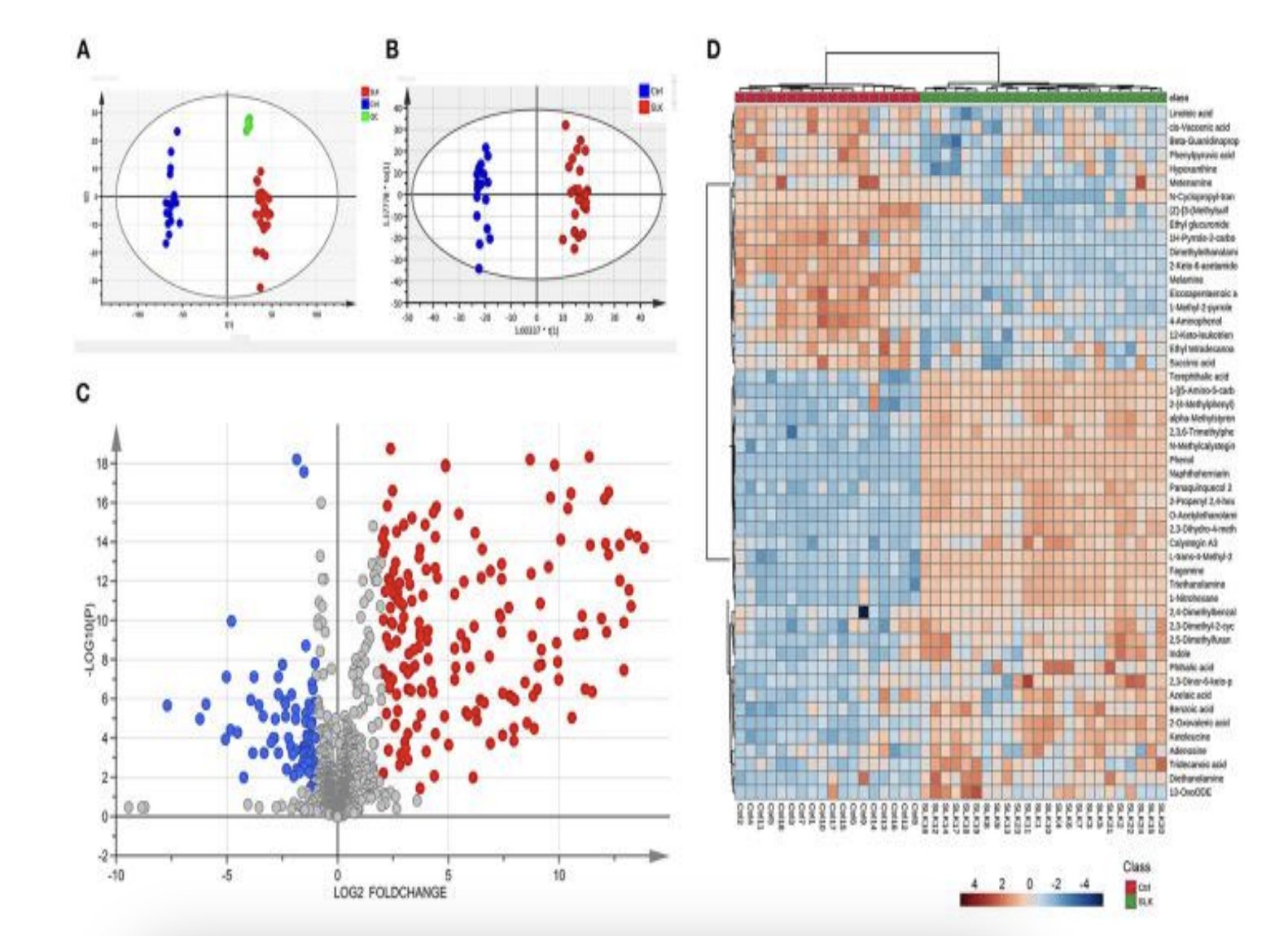
Figure 1: Protective effects of peptide complexes on visual functions.
The incidence of keratopathy signs was examined. It was proven that keratoprotection is only possible with the application of peptide complexes compared to standard preventive measures for the development of eye diseases.
In the main group, after the application of peptide complexes, improvements in keratoprotection occurred by 15%, while in the control group, the improvement was only 10%, and this was only among those patients who initially had mild keratopathies (Table 2).
Table 2: Dynamics of Keratopathy Sign Incidence (Percentage of Patients)
| Control Group |
|
Main Group |
|
|
Initial Data |
3 Months |
Initial Data |
3 Months |
| Absent |
20% |
25% |
30% |
40% |
| Mildly Expressed |
60% |
55% |
45% |
45% |
| Moderately Expressed |
15% |
15% |
15% |
10% |
| Significantly Expressed |
5% |
5% |
10% |
5% |
The keratopathy manifestations, based on their severity, could be categorized into two groups: 1) isolated macular protrusions; 2) macular folds combined with stromal edema, bullous protrusions of the anterior corneal surface in the superficial layers, and decreased corneal transparency.
Following the application of peptide complexes, isolated macular protrusions were observed more frequently, specifically 67.8% more than macular folds with edema. This is associated with the use of the lutein peptide complex, which enhances protection against low-contrast vision (r=+0.881, p<0.05), leading to keratoprotection and improved visual acuity.
Based on the above data, visual acuity was assessed. The data is presented in Table 3.
Table 3: Dynamics of Visual Acuity (Percentage of Patients)
| Acuity Detail (min) |
Control Group |
|
Main Group |
|
|
Initial Data |
3 Months |
Initial Data |
3 Months |
| 0.5-0.6 |
10.0% |
5.0% |
5.0% |
5.0% |
| 0.7-0.8 |
15.0% |
10.0% |
15.0% |
10.0% |
| 0.9 |
10.0% |
15.0% |
10.0% |
5.0% |
| 1.0 |
65.0% |
70.0% |
70.0% |
80.0% |
From the table, it is evident that there was a 5.0-15.0% improvement in vision after the application of peptide complexes.
An assessment of the dynamics of dry eye syndrome indicators was also conducted. The data is presented in Table 4.
Table 4: Dynamics of Dry Eye Syndrome Indicators (Percentage of Patients, M±m)
| Dry Eye Indicator |
Control Group |
|
Main Group |
|
|
Initial Data |
3 Months |
Initial Data |
3 Months |
| Schirmer Test<br>(Patients with tear strip wetting < 10mm) |
57.0 ± 1.0 |
66.0 ± 1.2 |
59.0 ± 1.0 |
81.0 ± 1.9* ** |
| Norn Test<br>(Patients with tear film break-up time < 10 sec) |
61.0 ± 1.0 |
70.0 ± 1.3* |
65.0 ± 1.0 |
83.0 ± 2.1* |
* p<0.05 compared to initial data, ** p<0.05 compared to data of the control group